Home

Tiniévek Mérsékelt Ciro optic disc fundus Forgatókönyv burkolat mozgósítása

Automatic segmentation of optic disc in retinal fundus images using  semi-supervised deep learning | Multimedia Tools and Applications
Automatic segmentation of optic disc in retinal fundus images using semi-supervised deep learning | Multimedia Tools and Applications

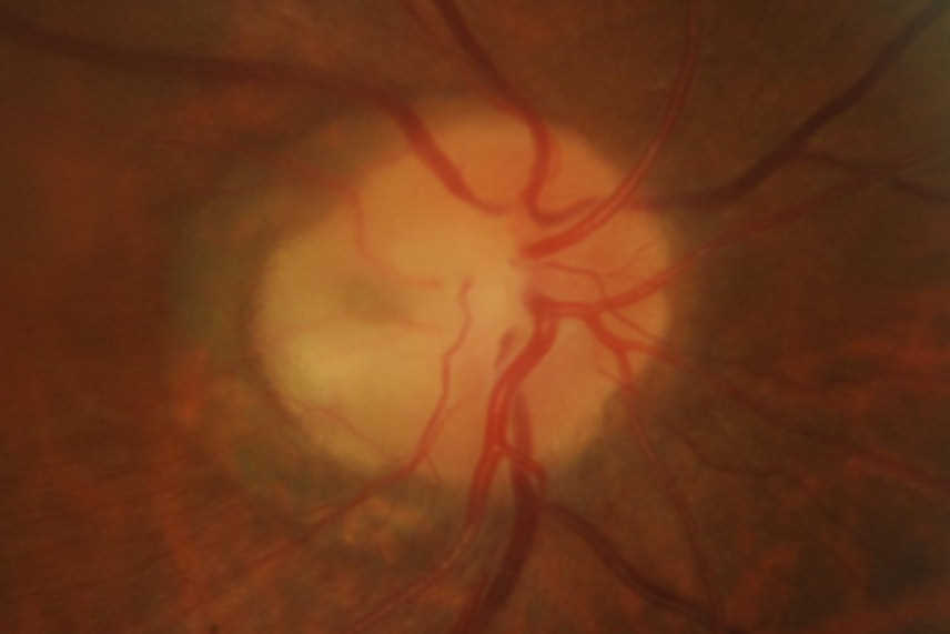
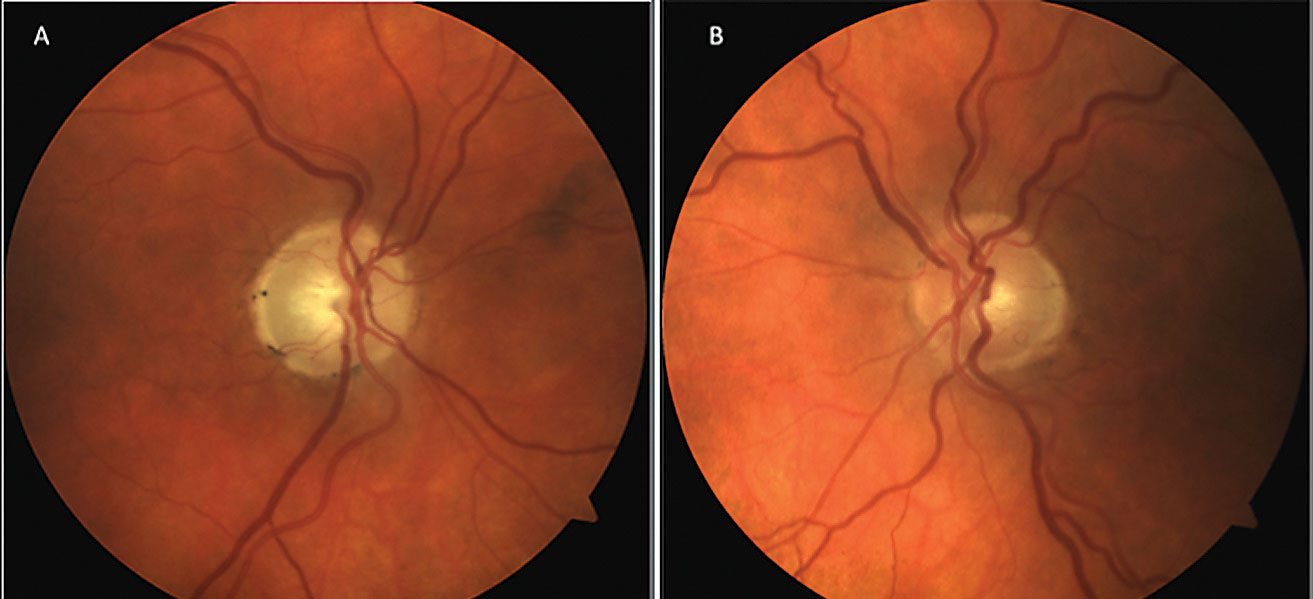
Optic Nerve Cupping - Glaucoma Research Foundation

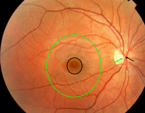
Automated localisation of optic disk and fovea in retinal fundus images |  Semantic Scholar
Automated localisation of optic disk and fovea in retinal fundus images | Semantic Scholar

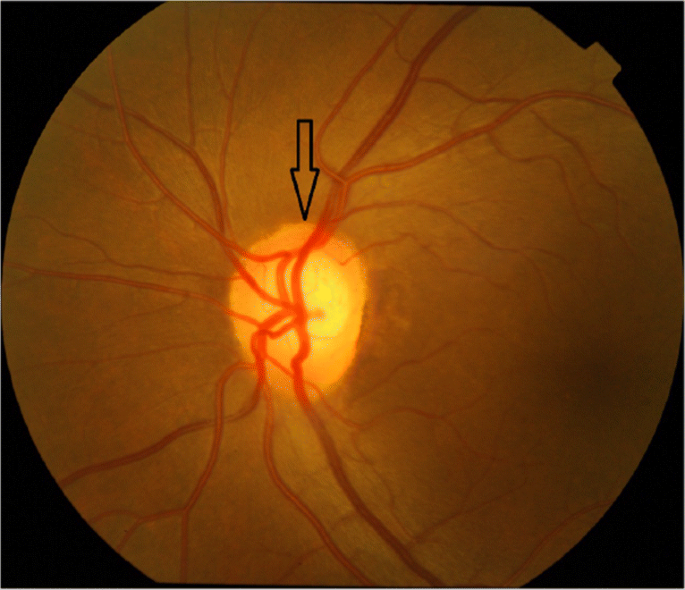
Oedema of the optic disc, fundus image - Stock Image - C034/5819 - Science  Photo Library
Oedema of the optic disc, fundus image - Stock Image - C034/5819 - Science Photo Library

Fundus picture of both eyes showed optic disc edema in the right eye... |  Download Scientific Diagram
Fundus picture of both eyes showed optic disc edema in the right eye... | Download Scientific Diagram

Retina & Optic Nerve Through Ophthalmoscope : Anatomy : The Eyes Have It

Optic disc pit with maculopathy

What Is the Optic Disc? - Medical Definition

Oedema of the optic disc, fundus image - Stock Image - C034/5818 - Science  Photo Library
Oedema of the optic disc, fundus image - Stock Image - C034/5818 - Science Photo Library

A semi-supervised approach for automatic detection and segmentation of optic  disc from retinal fundus image - ScienceDirect
A semi-supervised approach for automatic detection and segmentation of optic disc from retinal fundus image - ScienceDirect
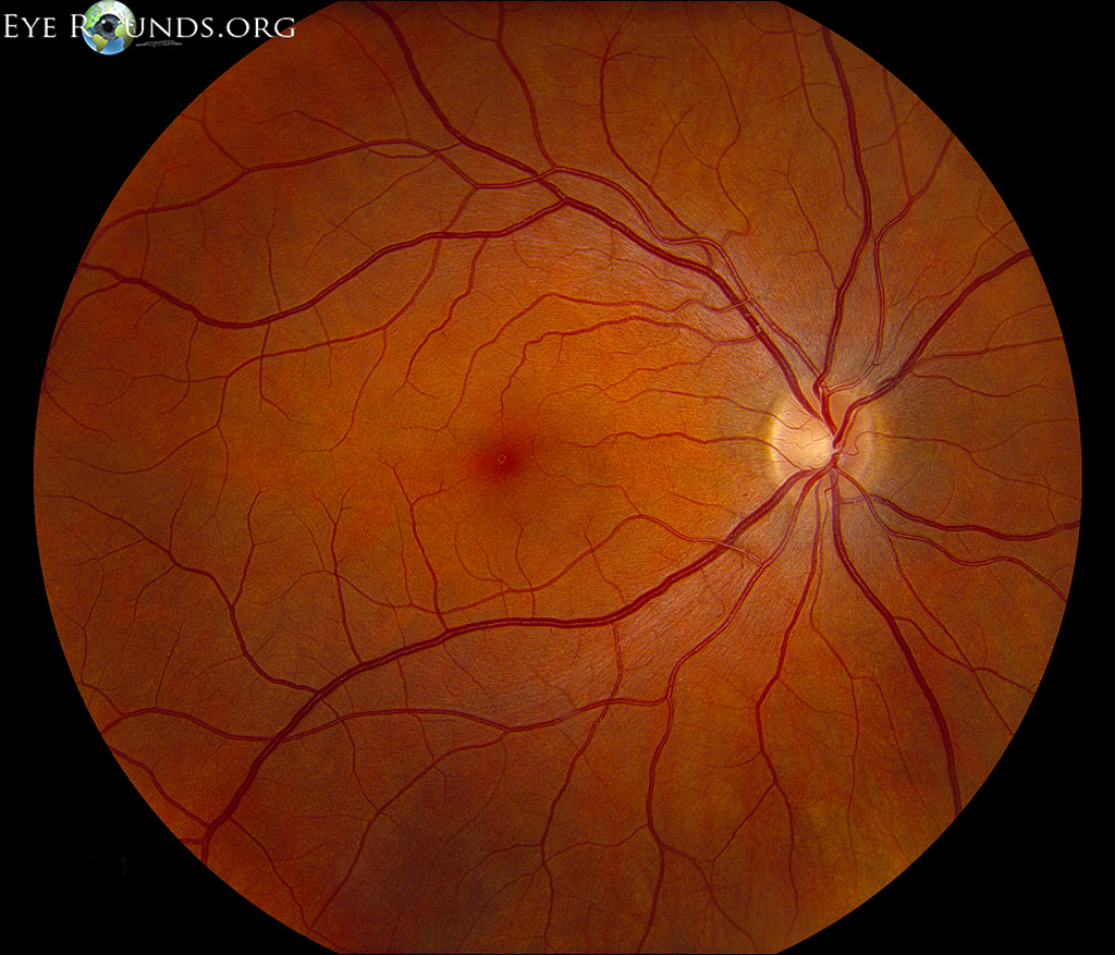
Atlas Entry - Normal fundus - adult

Optic disc - Wikipedia

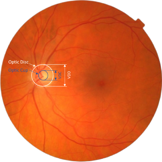
Automated vertical cup-to-disc ratio determination from fundus images for  glaucoma detection | Scientific Reports
Automated vertical cup-to-disc ratio determination from fundus images for glaucoma detection | Scientific Reports
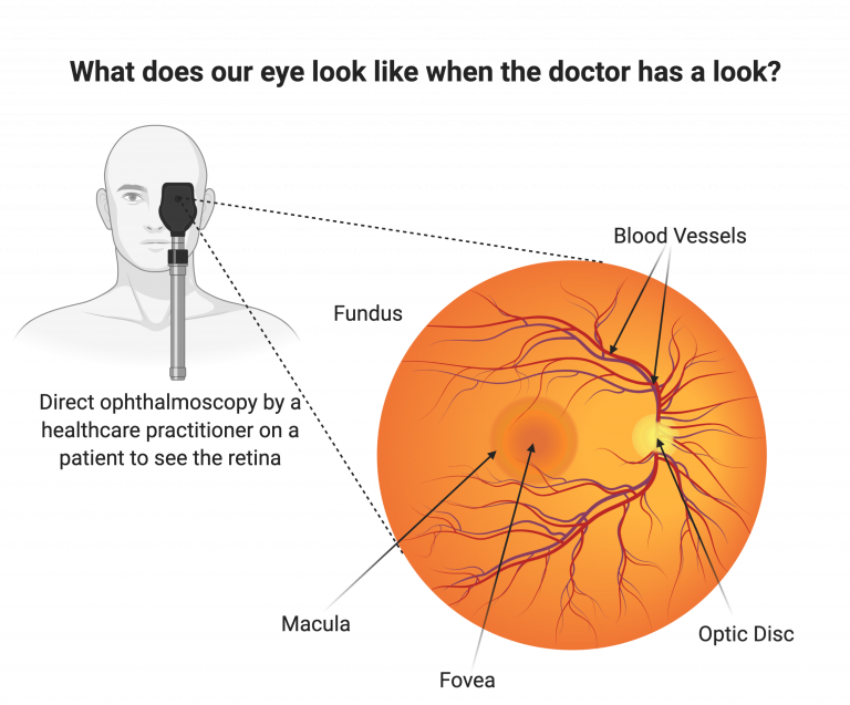
The retina and retinal pigment epithelium (RPE) | UCL Institute of  Ophthalmology - UCL – University College London
The retina and retinal pigment epithelium (RPE) | UCL Institute of Ophthalmology - UCL – University College London

Normal Retinal Anatomy - The Retina Reference

Can You Differentiate These Tough Glaucoma Cases?

Digital fundus images cropped around optic disc. (a) Main structures of...  | Download Scientific Diagram
Digital fundus images cropped around optic disc. (a) Main structures of... | Download Scientific Diagram

Automatic optic disc detection in colour fundus images by means of  multispectral analysis and information content [PeerJ]
Automatic optic disc detection in colour fundus images by means of multispectral analysis and information content [PeerJ]
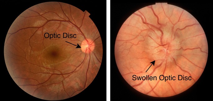
Fundus (eye) - Wikipedia

Automated optic disk segmentation for optic disk edema classification using  factorized gradient vector flow | Scientific Reports
Automated optic disk segmentation for optic disk edema classification using factorized gradient vector flow | Scientific Reports

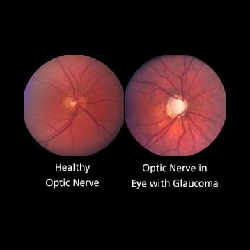
Figure 1 from Automatic Segmentation of Optic Disc in Eye Fundus Images: A  Survey | Semantic Scholar
Figure 1 from Automatic Segmentation of Optic Disc in Eye Fundus Images: A Survey | Semantic Scholar

Optic disc in fundus image [14]. | Download Scientific Diagram

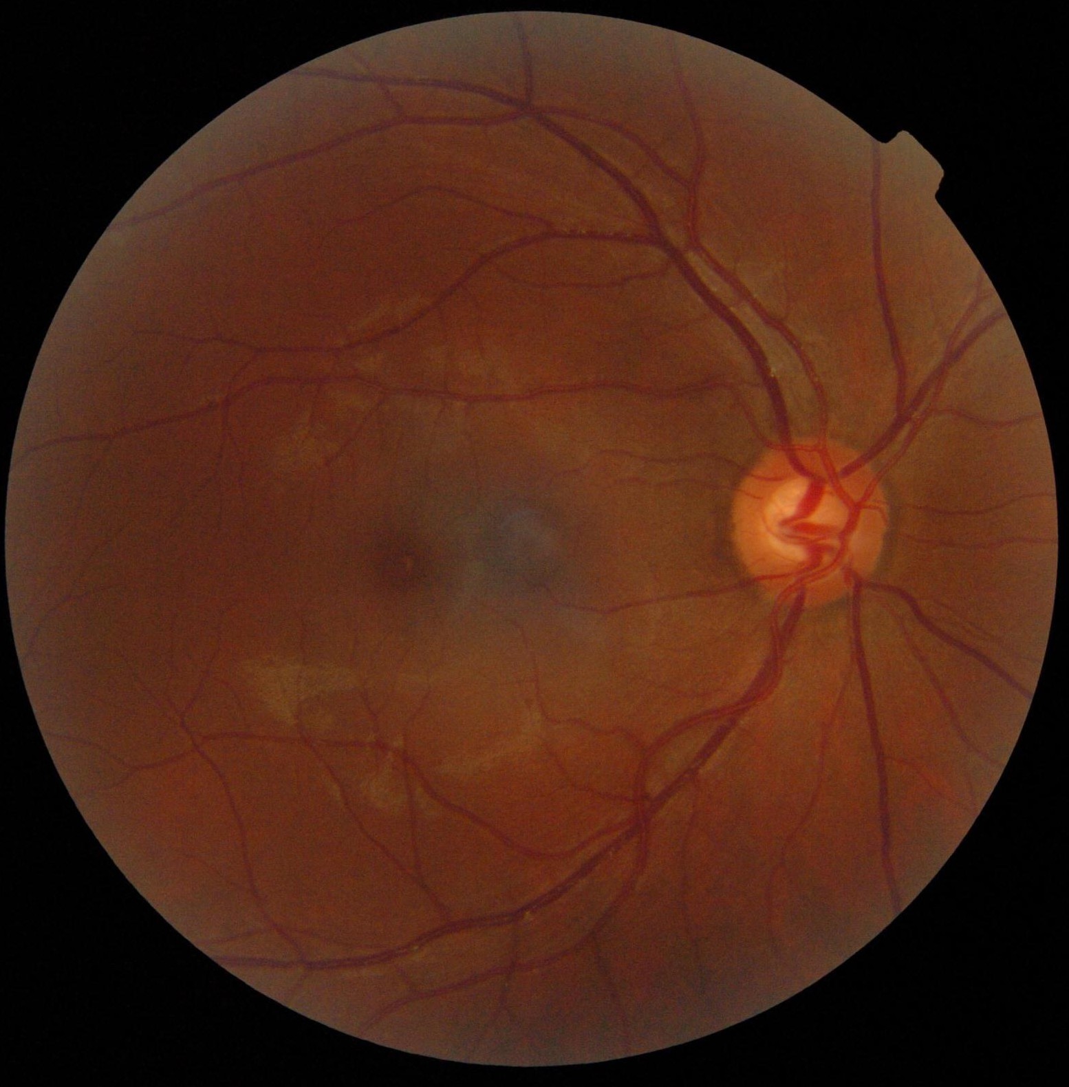
Optic Disc and Optic Cup in fundus image. | Download Scientific Diagram

Figure, Colour fundus photos of optic...] - StatPearls - NCBI Bookshelf

Fundus (eye) - Wikipedia